 
|
|
|
 |
 |
 |
|
|
|
|
For the simulation of soft tissues, our group has been developing an operative simulator that can be used in scleral buckling surgery for the treatment of retinal detachment of an eyeball.
For dynamic simulations, we perform an impact analysis that is used to analyze injuries caused by the expansion of an air bag and another analysis used in keratectomy to correct visual acuity. However, the entire eyeball has not been analyzed quantitatively and accurately. Therefore, we have been developing an analytical method to estimate the dynamic behavior during scleral buckling surgery for retinal detachment; in this technique, the eyeball is deformed by a sponge-shaped pad attached to the exterior of the eyeball or by a ring-shaped band that tightens the eyeball. The prognosis is greatly affected by the suture site or the extent of tightening. Previously, the amount of deformation and selection of the pad depended only on the experience and intuition of the doctor in charge. Since deformation progresses in a quasi-static manner, it is thought that selection of the shape of the silicon pad or the silicon band and the amount of ligation can be preoperatively simulated by finite element analysis.
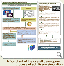
The system that we have been developing for the simulation of soft tissues is dhown in the figure below. The summary and overall flow of the development process are described in this figure. For further details, please refer to the linked web pages. |
|
| • Construction of a basic eyeball model
------------------------------------------------------------- |
As shown in the figure on the right-hand side, this study has been divided into two stages. The first stage is the initial construction of a basic eyeball model, and the second stage involves the construction of individual models using this basic model for the purpose of clinical application.
In the present project, we aim to develop the first stage of this study.
The construction of the basic eyeball model for the accurate simulation of an eyeball is described here. |
|
|
|
|
1. Digitization of the shape of an eyeball
The three-dimensional internal structure microscopy (3D-ISM) has been newly developed as a photographing device for simulation models; the eyeball was photographed using this technique.
Detailed information on the device is available at (--> Hideo Yokota’s home page). A human eyeball was obtained from the Eye Bank Association of America and used in this study. The photography conditions are available at (--> Sakiko Nakamura’s home page).
Based on this study, the detailed structure of an eyeball could be obtained at a resolution of 25 μm. This experiment was performed with the approval of the Faculty of Medicine, Toho University, and the ethical review committee of the Institute of Physical and Chemical Research.
|
2. Segmentation
The obtained image consisted of voxel data including all tissues, which were not divided into the data of each tissue. Therefore, we developed an automatic extraction method that uses image processing.
Detailed information on the automatic extraction method is available at (--> Tomoko Takemoto’s home page).
Each voxel was extracted manually by an operator who had anatomical knowledge, and a voxel model was constructed in which the eyeball was fractionated into its constitutive parts such as the sclera; cornea; crystalline lens; optic nerve; and retina, choroid, and the iris. The detailed construction method is available at (--> Nobunori Kakusho’s home page).
3. Measurement of dynamic characteristics
The stress-strain relationship of each tissue in the eyeball was measured by a tensile test. A measuring instrument was also developed.
Detailed results of the measurements are available at (--> Junko Sunaga’s home page).
4. Mesh generation
Based on the shape and physical property, a mesh for finite element analysis was generated by the mapped mesh method.
Detailed information on the mesh generation is available at (--> Shinobu Hirata’s home page).
5. A three-dimensional superelastic fine element analysis (FEM) program
As mentioned earlier, an eyeball is largely deformed by applying a force, and this deformation is nonlinear. Moreover, since hydrous materials such as the vitreous body exist in the eyeball, a coupled solid-fluid analysis is required. In order to perform a coupled analysis on an incompressible superelastic body and static fluid, a three-dimensional analysis program was developed.
Detailed information on the program is available at (--> Zhi-Gang Sun’s home page). |
|
|
| • Clinical application---------------------------------------------------------------- |
Using a basic simulation model that has been developed in the last 5 years, a clinically applicable simulator was developed.
In future, the basic eyeball model will be improved by including the parameters of an individual that are measured by nondestructive methods such as echography and MRI; this improved version will be used as an analytic model for clinical applications.
By using this analytical model, mesh generation for each patient will not be required, and the analytical model for each patient will be constructed in less time. Moreover, a database of typical diseases will be constructed; this will be used for establishing clinical criteria. |
|
| • Conclusion
---------------------------------------------------------------------- |
| As mentioned above, we have been developing a simulator that can be used for preoperative simulation of scleral buckling surgery for retinal detachment. In this project, we will develop a method to simulate tissues other than those of the eyeball based on the method used to construct the eyeball simulator. |
|
|
|
 |
|
|
